BulbiCAM - For the Neuro-Ophthalmologist What the OCT is for the Retina Specialist
 Often, life-threatening or serious conditions like intracranial tumours, aneurysms, neuropathies, and brain diseases affect the visual apparatus. This is often first suspected by an ophthalmologist as part of the neuro-ophthalmological workup.
The optic nerve is viewed, pupillary reactions verified, motility tested, movement modalities like smooth pursuit, saccades and nystagmus assessed, and visual field defects detected. Perimetries are frequently ordered, as well as OCT scans of the optic nerve. Many can make a diagnosis after a basic examination like this.
Still, referral to a neuro-ophthalmologist is often needed. Rare syndromes with unusual or hard to define symptoms may come into the picture. Eye, brain, muscles, nerves, and complex systemic diseases interplay. One must also be a neurologist. In many ways this makes neuro-ophthalmology. the most mentally challenging of all eye subspecialties.
Quantifying the exact extent of compromised motility, both in direction and degree, is time-consuming and can be hard to standardize. Different practitioners measure differently. This can make it difficult to determine progression over time. Determining the velocity and distance of saccades and nystagmus is something that can take years of practice to perfect.
Pupil assessment takes time – their size in darkness and light, cover/uncover, swinging flashlight, and indeed pupillary reaction tune and extent may be tedious to quantify. Also, here, exactness is an issue. This is one reason why many practitioners have pupillometers.
—
 I remember when my retina department first got an OCT. It was revolutionary. The diagnostic support it afforded us was invaluable. We could see, on an almost cellular level, the retina and how diseases affected it. Suddenly, we were able to make significantly faster and more accurate diagnoses. We could quantify edemas, and see how well our treatment worked. The changes brought by the OCT were far-reaching.
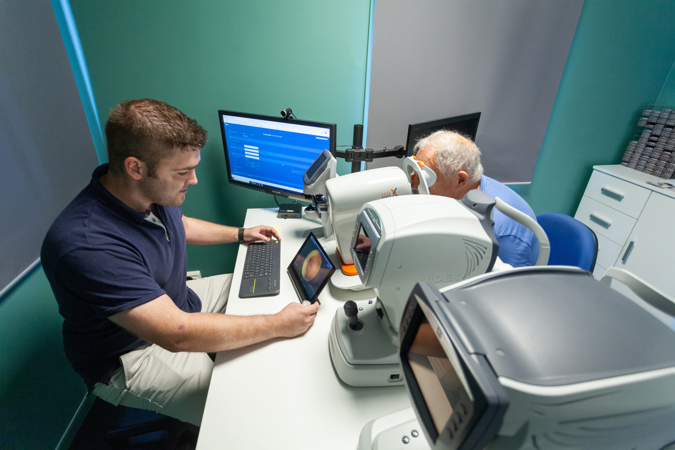
Enter BulbiCam – I believe it could be to neuro-ophthalmology what the OCT has been for the retina. How? It is built in a way that can film both eyes in motion, together with 3D VR screens that can display any colour image with a large field of view. The array of tests it can do is extensive – you can create virtually any visual stimulus, and observe the effect on pupils and motion – dynamically.
At an unrivalled (for non-research apparatus) 400 frames per second, it precisely measures the fastest of saccades – indeed this is the basis of a new form of glaucoma perimetry. Saccade velocity is affected earlier than the visual field. And the examination is done in less than a tenth of the time standard perimetry requires.
It can accurately quantify pupil size and reaction time indirectly and directly in darkness and light. Ptosis in varying directions of view may be measured. It can test color vision defects, contrast sensitivity, motility, and more. It does stereopsis.
Thus, we get great diagnostic support, requiring minimal operator intervention, and sparing us valuable time. Also, with its telemedicine capable software suite (BulbiHUB), it is easy to confer with my colleagues, sharing video, data, and images not only from BulbiCAM, but also from OCTs, fundus cameras, autorefractors, and other instruments.
Indeed, patients may be spared a visit to the neuro-ophthalmologist as these measurements may be done in my office, and easily be made available to neuro-ophthalmologists in other places. The suite has telemedicine built-in.
The great utility of BulbiCAM is what convinced me to join BulbiTech. I’m excited by its usefulness, and I’m persuaded that it will become a standard instrument, first for the neuro-ophthalmologist, and subsequently also for many others in eye care. Much like the OCT’s utility first was recognized by retina specialists, and later was adopted broadly.
