Please wait while flipbook is loading. For more related info, FAQs and issues please refer to DearFlip WordPress Flipbook Plugin Help documentation.
Eye Movement Perimetry
Both ocular and neurological disorders can lead to monocular or bilateral visual field defects. The intended use is to study effects of visual field defects on saccadic reaction times under various parameter settings, such as stimulus size or brightness.
Method: The eye movement perimetry tests are based on eye tracking and evaluates the eye’s ability to detect a peripheral stimulus while fixating on a central point. The test measures the saccadic reaction time, which is the time it takes for the eye to react and reach the peripheral stimulus. The test also includes visual tracing, and the results are presented in the output. The test presents different quantities of points depending on the program used, ranging from 16 to 60 points within 30 degrees of the fixation point.

16-point Test
Intended Use: To study the effects of large and/or pseudo defects associated with ocular motility limitations on saccadic reaction times under various settings. Ocular and neurological disorders can lead to ocular motility disorders, which can cause a pseudo visual field defect due to the inability of the eye to move towards the detected peripheral stimuli. The test evaluates 16 positions.
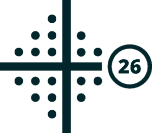
26-point Test
Intended Use: This test measures saccadic reaction times in areas of the visual field that are characteristic for glaucomatous visual field damage, such as superior and/or inferior paracentral and/or nasal defects. It simulates the 24-2 glaucoma screening pattern used in standard automated perimetry and evaluates 26 positions.
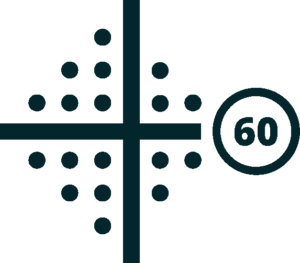
60-point Test
Intended Use: Measuring saccadic reaction times in a tightly spaced 60-position pattern can be used to study the effects of neurological disorders, such as lesions along the visual pathways, on delayed reaction times.
Scientific Publications on the Methodology
- Najiya Sundus Kadavath Meethal et al, Eye Movement Perimetry and Frequency Doubling Perimetry: clinical performance and patient preference during glaucoma screening Glaucoma, Published: 03 April 2019, Graefe’s Archive for Clinical and Experimental Ophthalmology volume 257, page
s1277–1287 (2019)
Please read here - Deepmala Mazumdar et al, Effect of Age, Sex, Stimulus Intensity, and Eccentricity on Saccadic Reaction Time in Eye Movement Perimetry, Author Affiliations & Notes Translational Vision Science & Technology July 2019, Vol.8, 13., doi:https://doi.org/10.1167/
tvst.8.4.13
Please read here - Deepmala Mazumdar et al, Comparison of saccadic reaction time between normal and glaucoma using an eye movement perimeter, Indian Journal of Ohthalmology, DOI: 10.4103/0301-4738.126182
Please read here - Deepmala Mazumdar et al, Saccadic reaction time in mirror image sectors across horizontal meridian in eye movement perimetry www.nature.com/
scientificreports, (2021) 11:2630 | https://doi.org/10.1038/ s41598-021-81762-y
Please read here - Deepmala Mazumdar et al, Visual Field Plots: A Comparison Study Between Standard Automated Perimetry and Eye Movement Perimetry, Journal of Glaucoma: May 2020 – Volume 29 – Issue 5 – p 351-361, doi: 10.1097/IJG.0000000000001477
Please read here - N. S. Kadavath Meethal et al, Development of a test grid using Eye Movement Perimetry for screening glaucomatous visual field defects, Graefes Arch Clin Exp Ophthalmol. 2018; 256(2): 371–379. Published online 2017 Dec 28. doi: 10.1007/s00417-017-
3872-x
Please read here

Dynamic Visual Acuity
Method: The best corrected visual acuity test aims to evaluate the eye’s ability to visualize and track a high-contrast moving stimulus that is gradually being reduced in size. The established thresholds achieved by the subject are then quantified and the measurements are represented in logMAR.
Intended Use: Determine the best achieved visual acuity values under best refractive conditions for the eye. Different pathologies, both ocular and neurological, can cause a decrease in visual acuity.

Contrast Sensitivity
Method: The contrast sensitivity test evaluates the eye’s ability to visualize and track a high contrast moving stimulus that is gradually being reduced in contrast levels and size. The established thresholds achieved by the subject are then quantified and the measurements are represented in logCon.
Intended Use: Determine the best achieved contrast sensitivity values under best refractive conditions for the eye and to study how certain pathologies of the retina affect the contrast sensitivity.

Smooth Pursuit Metrics
Method: The smooth pursuit test evaluates the eye’s ability to accurately track a visual target in a smooth controlled manner. The gain (%) variable, an expression of how close the eye is following the moving target, is measured.
Intended Use: Assessment of smooth eye pursuit capabilities. In some neurodegenerative disorders the eye might have reduced abilities to smoothly follow a target and tends to do catch-up saccades. The intended use is to detect and quantify eye pursuit dynamics.

Light Adaptation
Method: The macular light adaptation test aims to evaluate the central part of the retina’s adaptive mechanisms by showing the eye a moving stimulus in darkened conditions while periodically exposing the eye to a source of light with fixed or variable frequency. The highest threshold achieved by the eye while maintaining visibility of the stimulus is then quantified and themeasurements are represented in logCon or Hz.
Intended Use: To study the light-adaptive capability of the photoreceptors and retinal neurons in the foveal area, the central part of the retina.
Certain retinal disorders can affect the central retinal area with or without involvement of the fovea that is responsible for main vision.

Eyelid/Ptosis Metrics
Method: The ptosis test takes images of both eyes then quantifies the position of both upper and lower eyelids in relation to the cornea and pupil. The distance between the edges of the eyelids and the center of the pupil alongside with pupil diameters are measured.
Intended Use: Assessment of eyelids position in relation to the center of the eye and pupil diameter measurement. Ocular, neurological, endocrinological and other types of disorders can lead to malpositioning of one or both eyelids and/or asymmetry in pupils.

Fixation Stability Analysis
Method: The fixation test measures the fixation capabilities of an eye towards a stationary target before the occurrence of intrusive saccades, if any.
The test measures the span and peak velocity of eye movements in degrees.
Intended Use: Assessment of eye fixation capabilities. In some neurodegenerative disorders, the ability of an eye to fixate on an object is affected. The intended use is to detect and quantify fixation deviations.

Pro-Saccade Analytics
Method: The pro-saccade test aims to measure the saccadic latency, accuracy and peak velocity of fast eye movement, both short and long.
Intended Use: Assessment of saccadic eye movements. The intended use is to measure the values of fast eye movements. Saccade abnormalities are present in certain neurodegenerative disorders.
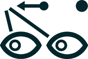
Anti-Saccade Analytics
Method: The anti-saccade test aims to measure the latency, accuracy and peak velocity of fast eye movement, both short and long by evaluating the gaze shift in the opposite direction from a presented target.
Intended Use: Assessment of anti-saccades. The intended use is to study the test subject’s ability to inhibit reflexive saccades.
Certain neurodegenerative disorders may cause a decrease in the ability to adequately control eye movement.

Memory Saccade Analytics
Method: The memory-saccade test aims to test subject’s transsaccadic memory by presenting a stimulus at a certain position, and then measuring the test subject’s capability to return to the same position after it disappears. Latency and accuracy of the gaze shift are measured.
Intended Use: Study of memory guided saccades. The intended use is to appreciate the test subject’s capability of short-term visual memory. Certain neurodegenerative conditions can affect short-term visual memory.

Express Nystagmus Analysis
Method: The nystagmography test is a video-based oculography method that measures involuntary eye movements in 9 cardinal directions at near (40 cm) and far (6 m) distances. The output displays eye movement amplitude, frequency and characteristics.
Intended Use: Study of nystagmus. The intended use is to video-record, detect, characterize and quantify involuntary eye movements. Certain ocular and neurological conditions lead to nystagmus.

Dynamic Pupillometry
Method: The pupil test evaluates pupil light responses in the form of changes in its diameter by exposing each eye to different screen brightnesses synchronously. Measurement output displays pupil diameters, pupil latency and peak velocity under various light conditions.
Intended Use: Assessment of pupillary light response. The intended use is to appreciate pupillary light responses and study their relationship to RAPD. Certain ocular and neurological disorders can cause monocular or bilateral pupillary defects in the form of response inadequacy.
Scientific Publications on the Methodology
Najiya S. K. Meethal et al, A haploscope based binocular pupillometer system to quantify the dynamics of direct and consensual Pupillary Light Reflex www.nature.com/scientificreports, Scientifc Reports | (2021) 11:21090 | https://doi.org/10.1038/s41598-021-00434-z
Please read here

RAPDlog Quantification
Method: Stimulates pupillary responses one eye at a time, alternating between the right and the left eye. With a bright light (pupil constriction) and exposes the pupil to darkness (pupil dilation), simultaneously or alternating.
Intended Use:
The RAPD test is used to detect a relative afferent pupil defect (RAPD), to detect differences between the two eyes in how they respond to light and dark environments. The test is intended to be used for detecting unilateral or asymmetrical signal transmission losses, such as can occur with heavy retinal opacities, unilateral retinopathies, glaucoma, or pathological pro- cesses in the optic nerve.

Saccade Metrics
Method: During a visual search task this test presents stimuli both horizontally and vertically. It is the patient’s task to search the presented stimuli and move the eyes towards it.
Intended Use: Saccade metrics are measurements used to study and analyze eye movements, specifically saccades. Saccades are rapid, jerky movements of the eyes that shift the line of sight between different points of interest in the visual field. Saccade metrics provide quantitative information about these
eye movements and can be used for various purposes, including: Research, diagnosis of ocular diseases, and evaluation of neurological diseases through signs of hyper- or hypometric eye movements.

Saccade Main Sequence
Method: The saccade main sequence graphically displays the relationship between the duration and amplitude of eye movements. The saccadic data areharvested from the visual field tests the patient has performed.
Intended Use: Using this test, researchers can gain a deeper understanding of the un- derlying neural processes and control mechanisms involved in saccades.
This information is crucial for investigating various aspects of oculomotor func- tion, such as the impact of brain regions, neurological disorders, injuries, and external factors on eye movement behavior.
